Ponerinae, Formicidae, Hymenoptera, Insecta, Arthropoda, Animalia
|
|
|
Additional Images:
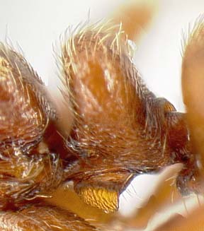
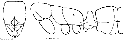
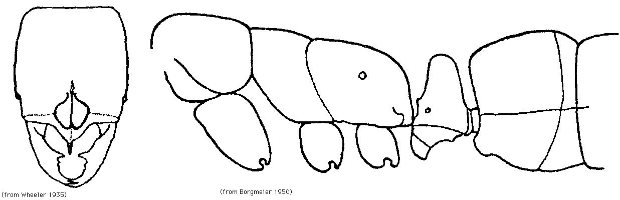
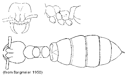
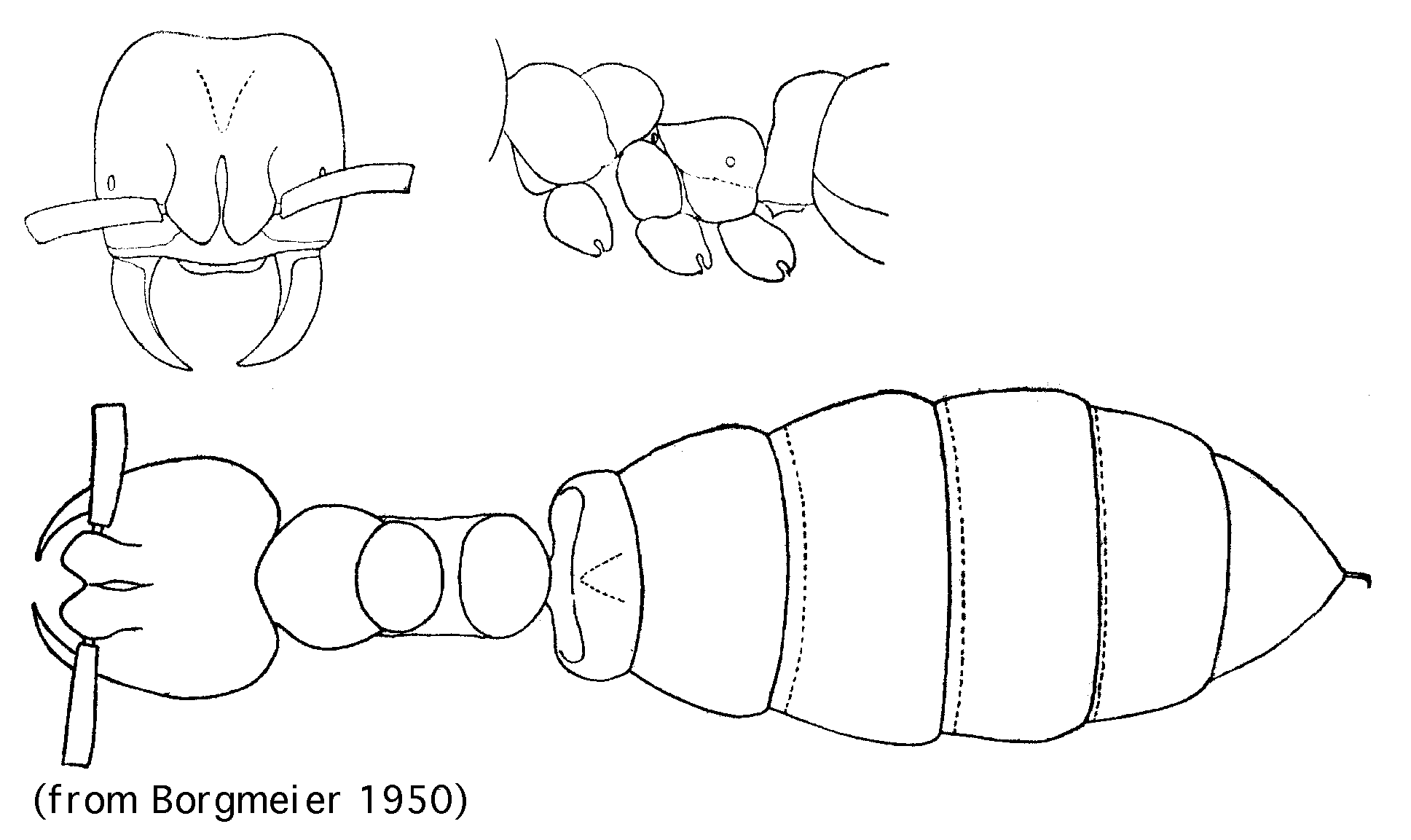
Worker, oblique view of petiole (small) (large); line drawings from Borgmeier (1950) and Wheeler (1935): worker face and lateral view (small) (large); queen (small) (large).
Range
Guatemala (type locality), Costa Rica (Central Valley, Atlantic slope).
Identification
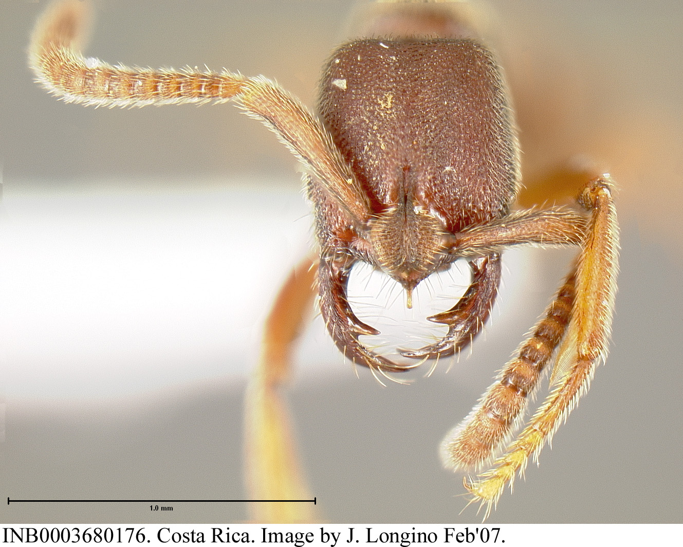
Anterior clypeal margin with spine, color brown, single faceted eye present.
Natural History
All Simopelta species are nomadic group raiders, convergent with Ecitoninae. See additional information under genus account.
Comments
Forel described pergandei from a broken specimen from Guatemala. Wheeler (1935) thoroughly redescribed pergandei, based on a "cotype" in Wheeler's possession, a Pergande specimen presumed to be part of the same series. Borgmeier (1950) described the queen based on collections from Costa Rica.
H. Schmidt collected multiple series of Simopelta pergandei, including at least two with colony queens, near San Jose, Costa Rica (Borgmeier 1950). Borgmeier stated that the material all came from the same site, with collection data La Caja, 8km from San Jose, H. Schmidt leg., 1940. I examined a recent topographical map of the vicinity of San Jose, and found a "Finca La Caja" a few kilometers along the road from San Jose to Juan Santamaria airport, at about 1000m elevation. Borgmeier illustrated both worker and queen from this material. The determination of the material as S. pergandei was based on a comparison of the workers with Forel's description, Wheeler's description, and Wheeler's figures. The specimens were examined by Brown, and he concurred with the determination (although he could not locate the "cotype" of pergandei, and surmised that it might be at AMNH).
Simopelta pergandei is described as having an anterior spine on the clypeus, small denticles between the second and third mandibular teeth, no mesosternal spine, red-brown color, and postpetiole broader than long in dorsal view.
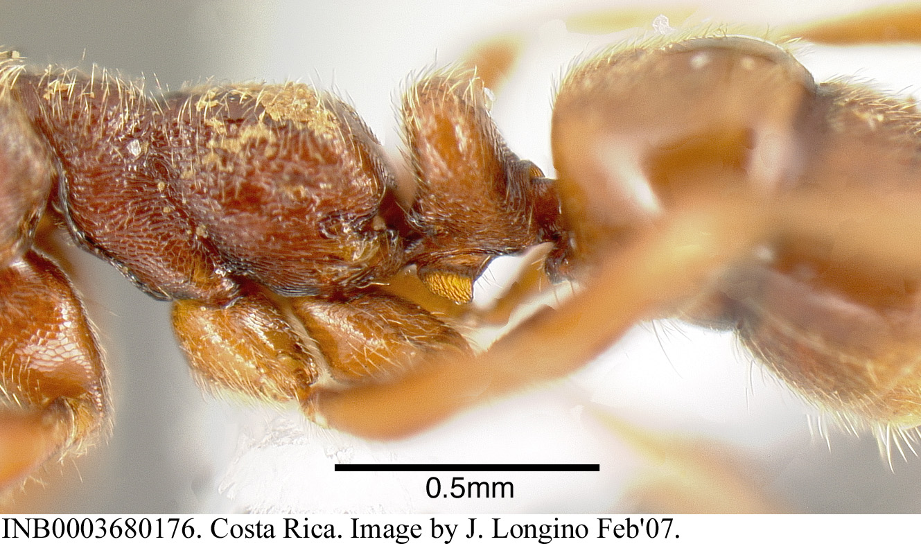
The illustrations of the queen of pergandei show the petiole broadly fused to the third abdominal segment. In contrast, the queens of JTL-001 and oculata have a typical constriction separating the petiole from the third abdominal segment.
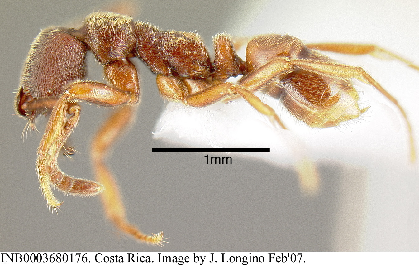
The TEAM Project, a biodiversity assessment and monitoring project sponsored by Conservation International, collected a single specimen of what I identify as S. pergandei. The images above are of this specimen. It was collected at 500m elevation in Braulio Carrillo National Park, a mature wet forest site near La Selva Biological Station. The worker was in a Winkler sample of sifted litter from the forest floor. The mandibles of this specimen differ from the descriptions of S. pergandei. Simopelta pergandei is described as having small denticles between the large basal tooth and the two large apical teeth, and the basal tooth is well separated from the apical teeth. On the TEAM specimen, the basal tooth is closer to the apical teeth, and there are no denticles between them.
Literature Cited
Borgmeier, T. 1950. A fêmea dichthadiiforme e os estádios evolutivos de Simopelta pergandei (Forel), e a descrição de S. bicolor, n. sp. (Hym. Formicidae). Rev. Entomol. (Rio de Janeiro) 21:369-380.
Forel, A. 1909. Ameisen aus Guatemala usw., Paraguay und Argentinien (Hym.). Deutsche Entomologische Zeitschrift 1909:239-269.
Wheeler, W. M. 1935. Ants of the genera Belonopelta Mayr and Simopelta Mann. Rev. Entomol. (Rio de Janeiro) 5:8-19.
Page author:
John T. Longino, The Evergreen State College, Olympia WA 98505 USA. longinoj@evergreen.edu
Last modified: 21 April 2007.